Treatment of OCD Treatment of OCD differs depending on the age of the patient and the severity of the injury. Studies suggest that younger patients with less severe OCD injury respond better to all forms of treatment.4, 13 The type of treatment used is dependant on the severity of the injury (see classification of OCD).3
Treatment of Type I Injury Patients with Type I injuries tend to respond well to non-operative treatment. This includes:
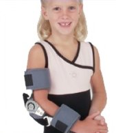
This rest period can last up to six months before the patient can return to their chosen activity. Follow up is carried out every three months with scans of the elbow to measure improvement.4,9,10,15,18 Patients should be counselled to consider changing their position in their chosen activity or possibly choosing another sport. Return to sport depends on whether pain is still present.4,5 |  Figure 9 Post-operative elbow brace |
Treatment of Type II Injury
Treatment is much the same as Type I injures. The patient should rest until the symptoms have settled and then start exercises to increase the movement and strength of the elbow.4,9,13,16
If after six months there is no improvement in symptoms then surgical treatment must be considered. Surgical treatments include:
- removal of loose bone fragments
- reattachment of loose bone fragments
- subchondral drilling (a procedure used to stimulate growth of new cartilage)
These can be done using open surgery, where an incision is made along the arm to gain access to the elbow, or by arthroscopy, where a number of small holes are made to allow instruments to be inserted to carry out the operation.4,10,13
The arm should be immbolised from five to twenty-one days to allow the bone to heal after surgery. After this time the patient is allowed to use the arm for everyday activities but not for any heavy lifting or sport. Physical therapy is often recommended to maintain elbow movement and strength.13,16
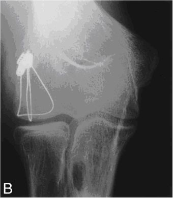
Follow up scans should be conducted every three months to check the progress of healing. Studies have shown that the material used for reattaching the fragment (screws and wires, see Figure 11) should be removed once healing has occurred. Leaving these materials in has been shown to increase the chances of damaging the surrounding cartilage.4,16
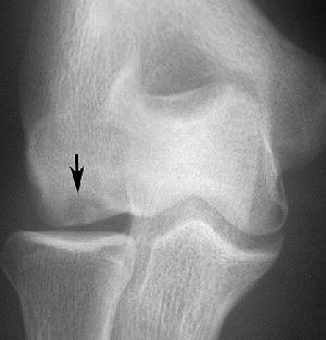
 Figure 10: X-Ray showing OCD of elbow |  Figure 11: X-ray showing elbow after surgery with wires holding bone fragment in place |
Treatment of Type III and Type IV Injury
These types of injuries have loose fragments in the joint and severe cartilage damage. In Type IV injuries the loose bone fragments also damage the head of the radius (bone of the forearm). Treatment for these injuries is similar to Type II injuries, where loose bodies are removed and in some cases a bone graft taken from another body part is attached to the humerus. Although, unlike Type I and Type II injuries, it is recommended that the patient should not return to sport.4
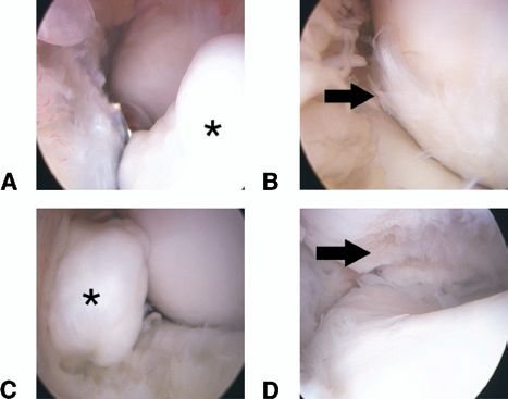
 | Figure 12: Shows images obtained during an arthroscopic procedure. The asterisks indicate loose bone fragments. The arrows indicate cartilage damage |
Prevention
Preventative measures are the best way to treat OCD. These include:17,18
- Education for coaches, parents and patients
- Limiting the number of throwing activities
- Using proper throwing technique
- Ensuring arm and elbow strength is adequate
Prognosis
Studies have suggested that if the disease is caught early then patients will respond better to treatment. More severe injuries generally do not heal as well as less severe injuries.4
- Type I injuries respond well to non-operative treatment with most patients returning to activity with similar levels of performance to before the injury.4,10,13,16
- Type II, Type III and Type IV injuries have conflicting evidence suggesting that some patients are able to return to the level of activity and performance and some suggesting that patients should not return to sport.4,13,16
OCD is linked to the onset of degenerative changes to the elbow joint later in life, such as degenerative joint disease and osteoarthritis. The future outcome is poor if degenerative changes to the joint are present as the cartilage on the joint surface will continue to wear away.4,10